Vithas Málaga introduces AI-powered MRI scanner to speed up examinations
The hospital has installed a Philips MR 5300 1.5T system that uses artificial intelligence to improve image acquisition, shorten scan times and make the experience more comfortable for patients.
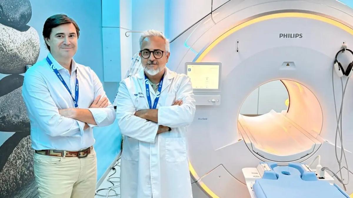

Vithas Málaga Hospital has introduced a new MRI scanner equipped with artificial intelligence, designed to speed up examinations while improving image quality and patient comfort.
The new system is a Philips MR 5300 1.5T, combining advanced image-processing technology with a wider bore and a system designed to significantly reduce the amount of helium needed for operation.
The scanner has already been integrated into the hospital’s daily activity and has completed its first 100 examinations, according to Vithas.
Artificial intelligence applied to MRI scanning
One of the main innovations is the use of artificial intelligence during image acquisition and reconstruction.
The technology helps optimise the information collected during the scan and produce high-quality images more efficiently.
For radiologists, this means having detailed images available for interpretation without unnecessarily extending certain imaging sequences.
The AI does not replace medical assessment. Instead, it works on the technical process used to capture and reconstruct the images.
Faster scans without sacrificing image quality
Reducing the duration of an MRI scan can benefit both radiology teams and patients.
During an MRI, patients usually need to remain still to prevent movement from affecting the images.
The shorter the time required to complete certain sequences, the easier it can be to maintain that position comfortably.
Vithas says the combination of artificial intelligence and advanced reconstruction tools allows examinations to be completed more quickly while maintaining high image quality.
Less time inside the scanner
For many patients, the length of an MRI is one of the least comfortable aspects of the procedure.
Patients must remain lying down and almost completely still while the scanner captures the different sequences.
Reducing this time can improve the experience, particularly for people who feel anxious, uncomfortable or find it difficult to stay in the same position for long periods.
The new system is designed to shorten that process whenever the type of examination allows it.
A wider 70-centimetre bore
The scanner also features a 70-centimetre bore.
The wider opening provides more space around the patient during the examination.
This can be particularly useful for people who experience anxiety or claustrophobia in narrower MRI systems.
It will not necessarily remove that sensation in every patient, but it does provide a physically more open environment.
Greater comfort can also help image quality
Patient comfort is not only about making the test more pleasant.
Movement is one of the main factors that can affect MRI image quality.
A patient who feels more comfortable may find it easier to remain still during the scan.
That means shorter examination times and a wider bore can also help the imaging process itself.
What does 1.5 tesla mean?
The new scanner has a magnetic field strength of 1.5 tesla.
Tesla is the unit used to measure the strength of the magnetic field generated by an MRI scanner.
1.5T systems are widely used in diagnostic imaging and can support a broad range of clinical examinations.
In this case, however, the innovation is not only about magnetic field strength.
Software, image acquisition, reconstruction and automation are also central to how efficiently the scanner performs.
AI works on acquisition and image reconstruction
Artificial intelligence can support several stages of the imaging process.
One is the acquisition of the data needed to create the scan.
Another is the reconstruction stage that follows.
Algorithms can help optimise the information collected so that clearer images can be produced using more efficient processes.
Radiologists then receive the resulting images and carry out the clinical interpretation.
Supporting the Diagnostic Imaging Unit
Dr Rafael Ramos de la Rosa, head of the Diagnostic Imaging Unit at Vithas Málaga, has highlighted the combination of speed and image quality.
The hospital says that obtaining sharp images in less time can make examinations more efficient and help specialists adapt protocols to the clinical needs of each patient.
The final medical interpretation remains the responsibility of the relevant specialist.
MRI does not use ionising radiation
MRI also has an important difference compared with some other imaging techniques.
It does not use X-rays or ionising radiation.
Instead, it relies on a strong magnetic field and radio waves to generate detailed images of the inside of the body.
For this reason, MRI is widely used to study many organs, tissues and anatomical structures.
Imaging different parts of the body
MRI can be used across many areas of diagnosis.
Common applications include neurological, musculoskeletal, abdominal, joint and soft-tissue examinations, among others.
The specific scan protocol depends on the part of the body being studied and the clinical reason for the examination.
The new technology allows imaging sequences to be adapted accordingly.
A more efficient approach to helium use
Another major feature of the new scanner is BlueSeal technology.
MRI systems need to keep certain parts of the magnet at extremely low temperatures.
Traditionally, this has required large amounts of liquid helium.
BlueSeal changes that approach by using a sealed circuit containing a much smaller quantity of helium.
Why MRI scanners need helium
Helium is used to cool the superconducting magnet inside an MRI system.
For the magnet to operate correctly, it must be kept at extremely low temperatures.
For decades, many MRI systems have relied on large volumes of helium to achieve this.
The challenge is that helium is a limited resource and its replacement also involves logistical and financial demands.
BlueSeal reduces the need for refilling
The system installed at Vithas keeps the helium in a sealed circuit.
This reduces losses and removes the need for the regular refilling associated with more traditional MRI systems.
As a result, the hospital can reduce its dependence on external helium supplies.
The manufacturer presents the technology as a way of making MRI operation more efficient.
Less reliance on a limited resource
Reducing helium use also has an environmental dimension.
Helium is a finite resource with important medical, scientific and industrial applications.
Using less of it can reduce the amount of material required throughout the lifetime of the scanner.
Vithas has highlighted this aspect as part of its wider technology upgrade.
Technology and sustainability in the same system
The new MRI scanner is designed around two main objectives.
The first is to improve diagnostic imaging through faster workflows and advanced image-processing tools.
The second is to reduce some of the resource demands associated with running the equipment.
BlueSeal is the main technology connecting those two goals.
The first 100 examinations have already been completed
Although the scanner was installed only recently, it is already fully integrated into the hospital’s activity.
According to Vithas, the system has completed its first 100 scans.
That confirms that it is already being used routinely with patients in the Diagnostic Imaging Unit.
Part of Vithas Málaga’s technology upgrade
The arrival of the Philips MR 5300 forms part of a wider process of technological renewal at Vithas Málaga.
The hospital is continuing to introduce equipment intended to improve diagnostic capabilities and make procedures more efficient.
In this case, the investment focuses on one of the most important tools for obtaining detailed images of internal structures.
AI supports diagnosis but does not replace doctors
Using artificial intelligence in an MRI scanner does not mean the machine makes the diagnosis on its own.
AI is mainly used as a technical support tool to optimise image acquisition and reconstruction.
Interpretation remains the responsibility of healthcare professionals.
Radiologists analyse the images while taking into account the reason for the examination, the patient’s clinical history and any other relevant information.
The most noticeable changes for patients
Some of the technology operates behind the scenes and may not be obvious to patients.
However, two improvements are likely to be particularly noticeable.
The first is the wider 70-centimetre bore.
The second is the possibility of completing some examinations in less time.
Both are aimed at making MRI scanning easier to tolerate.
Claustrophobia remains a common challenge
Some people experience anxiety when entering an MRI scanner.
This can be particularly noticeable during longer examinations or when there is little space around the body.
A wider bore creates a greater sense of openness.
If the scan can also be completed more quickly, patients spend less time inside the machine.
More comfort for different types of patients
The wider opening may be useful not only for people with claustrophobia.
It can also make positioning easier for some patients and provide additional space during certain examinations.
Each scan still has its own technical requirements.
The Diagnostic Imaging team determines the most appropriate protocol in each case.
Less movement can mean better images
Movement is one of the main factors that can reduce the quality of an MRI scan.
If a patient changes position during a sequence, some images may become blurred and need to be repeated.
Shorter examinations can make it easier to remain still.
That can improve efficiency and help avoid unnecessary repetition where possible.
Málaga adds another advanced imaging system
The new scanner expands the diagnostic technology available in Málaga’s private healthcare sector.
MRI systems are evolving not only through stronger magnets but increasingly through software.
Advanced processing, automation and artificial intelligence are becoming major areas of development.
The Philips MR 5300 installed at Vithas Málaga is part of that wider technological shift.
Speed, comfort and efficiency are the main changes
The new system can be summarised through three main improvements.
First, artificial intelligence is used to optimise imaging and shorten examinations.
Second, the 70-centimetre bore provides patients with more space.
And third, BlueSeal technology significantly reduces helium requirements.
They are different innovations, but all are aimed at making MRI examinations more efficient for both patients and the hospital.
A new stage for the Diagnostic Imaging Unit
After its first 100 examinations, the scanner is now part of normal daily activity at Vithas Málaga.
Its introduction updates one of the hospital’s key diagnostic imaging technologies.
Artificial intelligence is being used here not as a replacement for medical professionals, but as a tool for obtaining high-quality information more efficiently.
For patients, however, the most noticeable change may be simpler: spending less time inside a wider MRI scanner during a test that can often cause some anxiety.

Latest news