Costa del Sol Hospital introduces MRI-guided breast biopsy for complex cases
The Marbella hospital has added a minimally invasive technique that allows doctors to sample lesions that cannot be located by mammography or ultrasound, improving diagnostic precision and treatment planning.
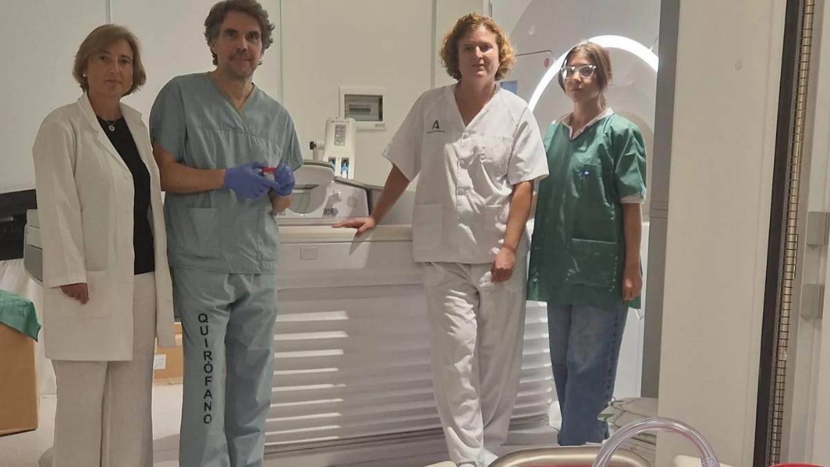

Costa del Sol University Hospital in Marbella has introduced MRI-guided breast biopsy within its Radiology Department, adding a new diagnostic option for patients whose suspicious lesions can only be seen on magnetic resonance imaging.
The new procedure expands the capabilities of the hospital’s Breast Unit and makes it possible to obtain tissue samples with greater precision without immediately resorting to exploratory surgery.
It is particularly useful in more complex cases where digital mammography and ultrasound are unable to identify an abnormality that is visible on MRI.
A technique for lesions only visible on MRI
The main feature of the procedure is the type of lesion it is designed to investigate.
It does not replace standard breast biopsies and is not intended for routine use in every patient.
Its purpose is to obtain tissue from suspicious abnormalities detected exclusively by MRI.
These lesions can be very small and completely invisible on other imaging tests.
MRI makes it possible to locate them and guide the biopsy system accurately towards the area that needs to be analysed.
Some cases previously required exploratory surgery
The introduction of the technique changes the approach to cases where a lesion cannot be identified using ultrasound or mammography.
Until now, when an abnormality was visible only on MRI, specialists could sometimes need to perform exploratory surgery to obtain a tissue sample.
MRI-guided biopsy provides a less invasive alternative.
It allows the suspicious tissue to be reached through the Radiology Department without initially requiring an operation in theatre.
How MRI-guided breast biopsy works
The procedure combines MRI imaging with a vacuum-assisted biopsy system.
During the examination, MRI is used to identify the exact position of the lesion.
Once located, the medical team guides a needle into the area and removes small tissue samples for analysis by Pathology.
The precision of the imaging is especially important when the abnormality measures only a few millimetres.
A vacuum-assisted biopsy system
Costa del Sol Hospital uses a technique known as vacuum-assisted biopsy, or VAB.
The device uses a needle connected to a vacuum system that allows several tissue samples to be collected continuously.
One key difference compared with a conventional core needle biopsy is that the needle does not need to be repeatedly removed and reinserted for every sample.
This makes it possible to obtain several specimens from the same position.
Several samples through a single access point
The ability to collect multiple tissue fragments without repeatedly puncturing the area is one of the main features of the technique.
The aim is to obtain enough material for specialists to determine the nature of the lesion as accurately as possible.
The samples are then examined to confirm or rule out malignant cells.
This means a diagnosis can be reached without having to remove the entire suspicious area at the outset.
An outpatient procedure lasting 30 to 45 minutes
Another advantage is that the procedure does not require hospital admission.
The biopsy is carried out as an outpatient procedure under local anaesthetic.
It takes approximately 30 to 45 minutes.
Once the procedure has been completed and the necessary checks have been made, the patient can return home the same day.
A small marker is left in place
After the samples have been taken, a small marker clip is placed at the site of the lesion.
This has a practical purpose.
If surgery or another treatment is later required, the marker helps doctors identify precisely where the biopsied area was located.
The clip is very small and remains in place as a reference for future imaging or treatment.
Helping surgeons plan treatment more accurately
The usefulness of the procedure goes beyond confirming a diagnosis.
Information obtained through MRI and biopsy can also help determine the true extent of the lesion.
This is particularly important if the patient later needs surgery.
Knowing the exact area involved can help surgeons plan the operation and decide how much tissue needs to be removed.
The aim is to preserve as much healthy tissue as possible
A more accurate definition of the disease can help tailor treatment to the individual patient.
When surgery is required, having a clearer map of the lesion may make it easier to remove the affected tissue while preserving as much healthy breast tissue as possible.
This level of precision is especially important in breast-conserving surgery.
Better information before surgery can therefore support more detailed multidisciplinary planning.
Particularly useful for women with dense breasts
MRI-guided biopsy can be especially useful in women with dense breast tissue.
In these cases, certain abnormalities may be more difficult to identify using conventional imaging techniques.
MRI can provide additional information and reveal suspicious areas that are not visible on other tests.
When that happens, MRI-guided biopsy makes it possible to take a sample directly from the area concerned.
Also relevant for women at high genetic risk
Another group that may benefit includes women considered to be at high genetic risk of developing breast cancer.
These patients may undergo enhanced surveillance programmes in which MRI plays an important role.
If one of these scans identifies a lesion that is visible only on MRI, the new technique allows it to be investigated without immediately turning to surgery.
Useful for patients who already have a cancer diagnosis
The procedure also has applications in women who have already been diagnosed with breast cancer.
In some cases, doctors need to determine whether there are additional tumour sites within the same breast.
This is known as multifocal disease.
MRI can reveal small additional lesions that were not detected by other imaging techniques.
Biopsy can then determine whether these areas are also malignant.
It can also assess the opposite breast
MRI may also identify a suspicious abnormality in the opposite breast.
Before changing treatment or planning more extensive surgery, it is important to know whether that second area is genuinely malignant.
A targeted biopsy can provide this information using a minimally invasive procedure.
Around 30 women could benefit each year
Costa del Sol University Hospital estimates that around 30 patients per year could benefit from the new service.
The relatively small number reflects how specific the technique is.
Most breast lesions will continue to be investigated using conventional methods.
MRI-guided biopsy is reserved for cases in which other imaging techniques cannot locate the suspicious area.
An advance in pre-surgical diagnosis
Introducing the procedure gives specialists more options before deciding on surgical treatment.
The medical team can obtain tissue from complex lesions without using an operating theatre as the first diagnostic step.
This may help shorten the pathway between detecting an abnormality and making a treatment decision.
It can also reduce some of the uncertainty associated with waiting for multiple tests.
Breast radiology remains central to diagnosis
The Radiology Department plays a major role in detecting and characterising breast lesions.
Mammography, ultrasound and MRI each provide different and often complementary information.
The choice of test depends on the patient, the type of lesion and the information specialists need.
With the new biopsy service, Costa del Sol Hospital can now complete the investigation of certain MRI-only abnormalities within the same centre.
A multidisciplinary approach
Diagnosing and treating breast cancer requires close cooperation between different specialists.
Radiologists, pathologists, surgeons, oncologists and nursing teams all contribute to the assessment of each case.
MRI-guided biopsy becomes another part of that coordinated pathway.
The biopsy results are then combined with the rest of the clinical and imaging information to determine the most appropriate treatment strategy.
Costa del Sol Hospital expands its clinical services
With this addition, Costa del Sol University Hospital joins other Andalusian hospitals already offering the technique.
The Andalusian regional government says the development improves access to advanced diagnostic procedures within the public healthcare system.
For patients in the hospital’s catchment area, it also means that this specialised procedure can now be carried out at their local reference hospital.
Minimally invasive does not mean everyone needs it
The introduction of the technique does not mean it will be used in every breast assessment.
Mammography and ultrasound remain essential diagnostic tools.
MRI and MRI-guided biopsy are used in specific circumstances where they can provide additional information.
The medical team will decide which test is appropriate in each individual case.
A new option for highly complex cases
The real importance of the new service lies in addressing a very specific diagnostic challenge.
When a suspicious finding cannot be located using standard imaging, obtaining a tissue sample can be difficult.
MRI-guided biopsy turns the MRI image itself into a precise roadmap for reaching that area and analysing it.
In doing so, Costa del Sol Hospital expands its ability to provide more accurate diagnosis and more precisely planned treatment for complex breast cases.

Latest news